C O A C H I N G
Gaba
Introduction
Neurons are what we call the cells inside the brain that produce and respond to neurotransmitters, and other chemical messengers. Most neurons have a storage of neurotransmitters contained inside of them, inside of vesicles. When a neuron is at “rest”, meaning it is not releasing its stored neurotransmitters, it is referred to as being inhibited. In order to excite a neuron it must surpass a voltage threshold, which allows for different ions, specifically calcium, to enter into the cell and allow vesicles containing neurotransmitters to fuse with the cellular membrane and release the stored neurotransmitters. The neurotransmitters are released out of the neuron into the synaptic space where they can bind to a receptor located on another neuron. The synaptic space is simply the space between two communicating neurons. When the voltage surpasses a specific threshold, the vesicle is capable of fusing into the cellular membrane and releasing the neurotransmitters stored inside. To be slightly less abstract, the resting potential of a neuron is estimated to be around -70 millivolts. This means, that inside the neuron is 70 millivolts lower than the environment outside of the neuron. These numbers are quantified according to the charge of all the molecules within a neuron. It is estimated that when a neuron surpasses the voltage of 50 millivolts, it becomes capable of releasing the stored neurotransmitters.
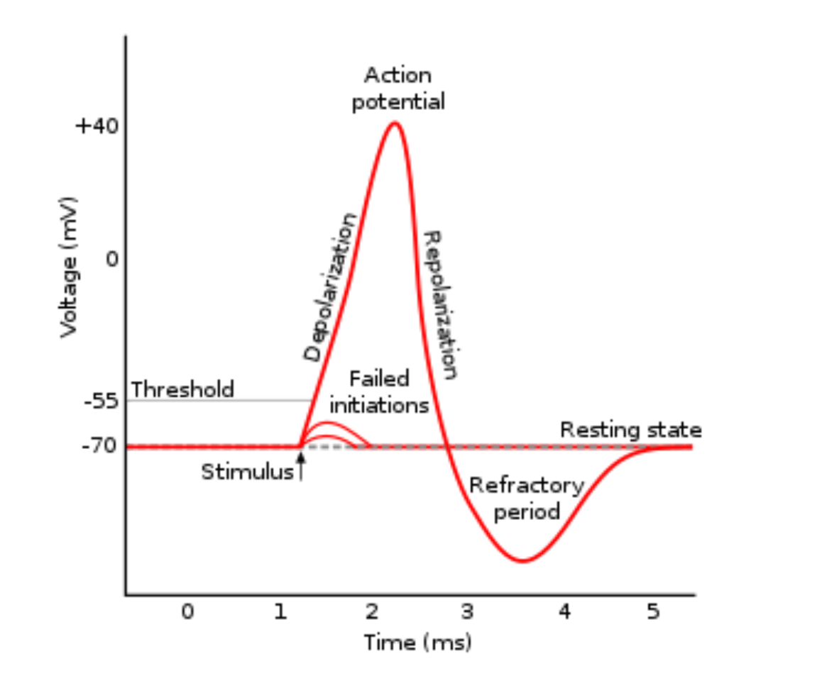
Within the nervous system, we have broad classes of neurons referred to as excitatory and inhibitory neurotransmitters. Excitatory neurotransmitters are neurotransmitters that assist the neuron in reaching its voltage threshold by increasing the amount of positively charged molecules that enter the neuron. Conversely, inhibitory neurotransmitters typically cause positively charged ions to leave the neuron, and negatively charged ions to enter, thus decreasing the voltage of the neuron and making it more difficult to reach its voltage threshold.
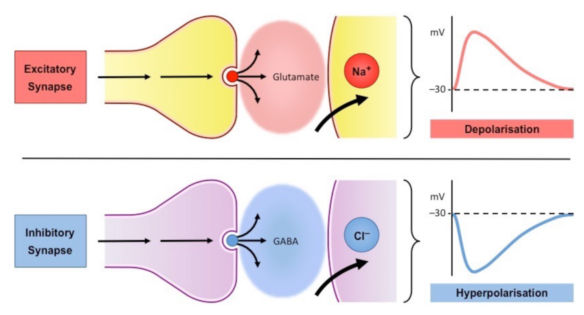
For instance, GABA will lower the voltage of a neuron, therefore inhibiting it from reaching the voltage required for neurotransmitter release. Conversely, glutamate is considered an excitatory neurotransmitter, and it increases the voltage of a neuron enough to secrete the neurotransmitter. Before a neuron actually releases a neurotransmitter, there is a multitude of events that occur in order for the vesicle containing the neurotransmitter to fuse to the membrane of the neuron and release the neurotransmitter out of the neuron. Luckily, one can actually witness what the excitation of a neuron looks like as I discuss the release of GABA, then I will subsequently describe how and why GABA is referred to as the inhibitory neurotransmitter.
Production and storage
GABA is secreted by neurons located in the central nervous system. The neurons in the spinal cord that store and release gaba are referred to as the presynaptic GABAerigic neurons. These neurons store and release GABA into the area known as the synapse. Remember that the synapse is simply the space between two communicating neurons. The neuron that is inhibited by GABA is referred to as the postsynaptic neuron.
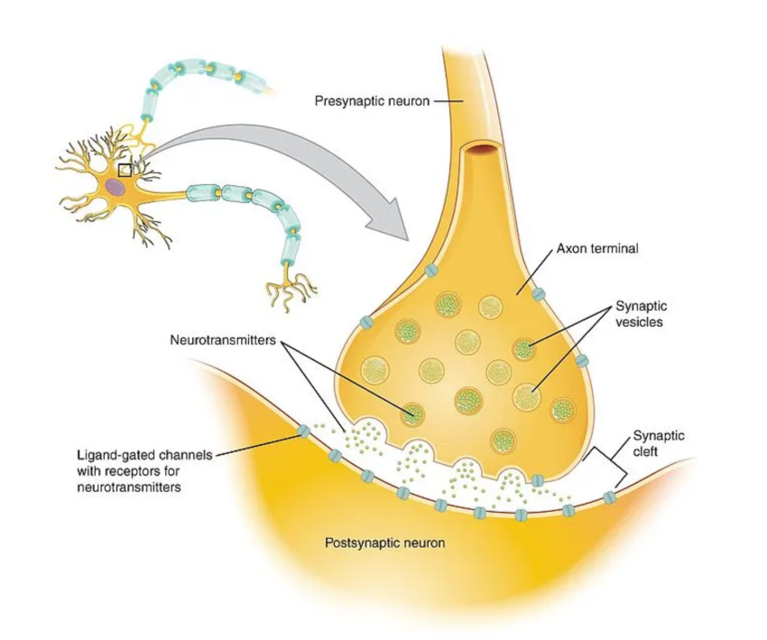
Before we get into the effects of GABA, let's talk about how we get GABA into the secretory vesicle within the presynaptic neuron in the first place. Interestingly, GABA isn't actually able to be uptaken into the neuron in order to be stored. Thus, neurons need to uptake a different molecule out of the bloodstream called glutamine, which is eventually converted to GABA. Glutamine can enter through a group of amino acid transporters called sodium coupled neutral amino acid transporters (SNATs) (1). Glutamine is then converted into glutamate via the glutaminase enzyme. Then, via another enzyme called glutamate decarboxylase, we can convert glutamate into GABA. A very important caveat is that the conversion of glutamate to GABA is dependent upon vitamin B6. Thus, a deficiency in vitamin B6 may alter the balance of glutamate to GABA, in favor of glutamate. It is postulated that the excessive amount of glutamate, which is an excitatory neurotransmitter, is a very strong driver of cognitive deficits such as ADHD, anxiety, and even sleep disturbances (2-4)
Release of GABA
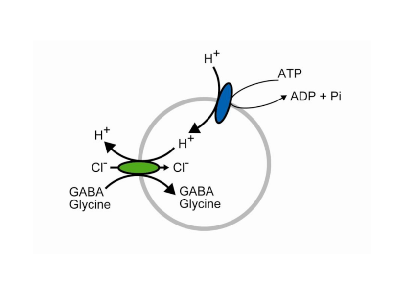
Once we have synthesized GABA, it is stored in what are called V-GAT vesicles. The uptake of GABA into these vesicles is quite interesting. It occurs through a hydrogen ATPase protein on the membrane of the vesicle. This protein utilizes ATP, removing hydrogen from ATP. This hydrogen is used as a substrate for the antiporter on the me membrane reffered to as V-GAT. An anti-porter is simply a protein channel that will update one molecule while subsequently releasing a stored molecule. This anti-porter will release the stored hydrogen back out of the vesicle and uptake the free GABA that was created originally from glutamine.
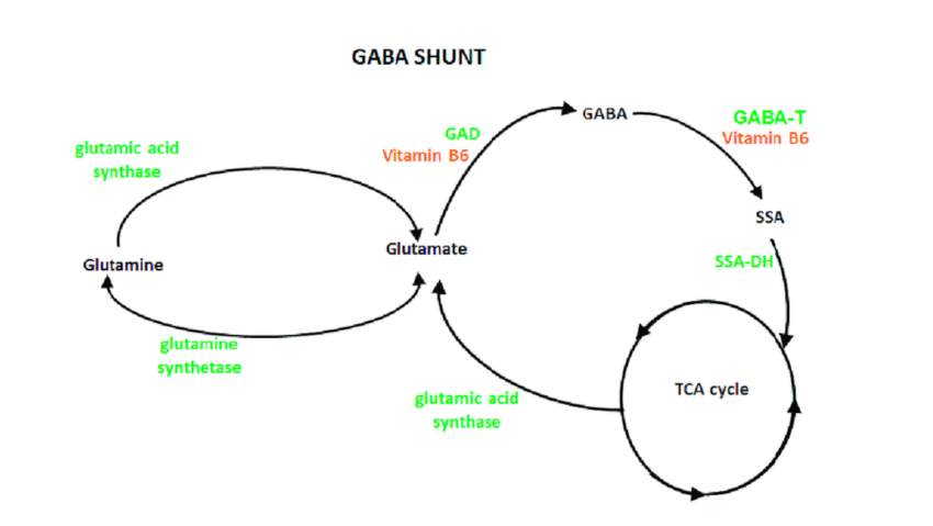
There are 2 very important nutritional caveats here. Glutamine is an amino acid derived from protein, thus an insufficient intake of dietary protein certainly has the potential to dysregulate adequate glutamate and gaba production. Moreover, as mentioned vitamin B6 plays a crucial role in the conversion of glutamate to GABA, therefore B6 could potentially being underlying deficiency and driving symptoms of ADHD and anxiety.
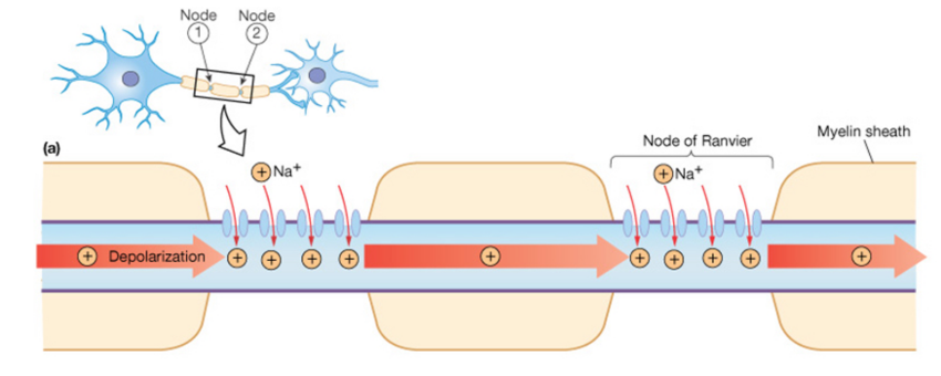
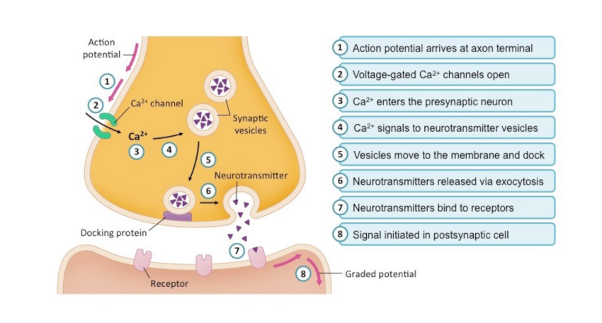
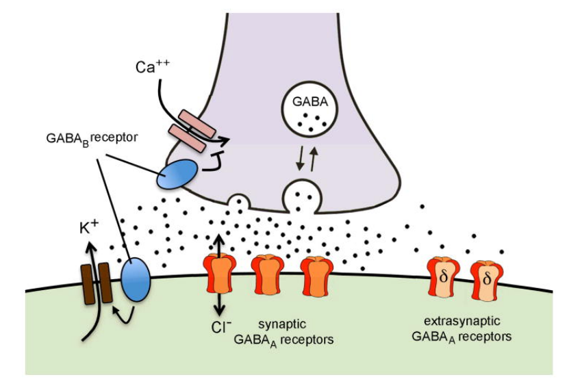
In order to use GABA, it must be released from the V-GAT vesile, into the synapse. This is stimulated through a series of mechanisms that increase the concentration of positively charged molecules inside of the GABAergic neurons, thus causing the neuron to surpass the voltage required to release the stored GABA. This is referred to as excitation, and it said exactly the opposite of what GABA does! This process begins when sodium channels located along the axon of the neuron open. The sodium molecules are positively charged, and they increase the voltage of the neuron. This will stimulate voltage gated calcium channels to change shape. As the name implies, the activity of these channels is dictated by the voltage of the neuron. When the neuron reaches a specific voltage these calcium channels change shape and allow calcium influx into the neuron.
Calcium is the substrate that allows the vesicle containing GABA to fuse the plasma membrane and release the GABA stored inside of the vesicle. This is mediated by proteins present on the vesicle and on the plasma membrane referred to as SNARE proteins. Calcium allows these proteins to attach to one another, essentially fusing the vesicle into the membrane, and release the stored neurotransmitter, which in this case is GABA.
Calcium is necessary for allowing the vesicle to fuse to the membrane of the neuron, and release GABA outside of the neuron. Calcium allows for the interaction of proteins called SNARE proteins, located on the membrane of the vesicle, and the membrane of the neuron to bind to one another and fuse the vesicle to the membrane.
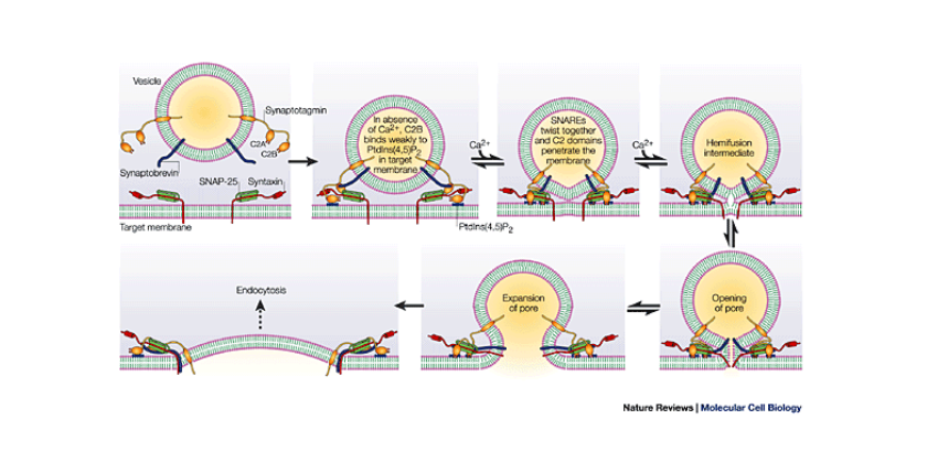
If you're a nerd like me, here is the cellular physiology of this process:
On the membrane of the vesicle, there is a protein called synaptobrevin. Also, on the membrane of the vesicle is another protein called synaptotagmin. On the membrane of the neuron, where the vesicle will dock, there’s a protein called syntaxin. Additionally, on the membrane, there is a protein called SNAP-25. Simply, these three proteins form a complex that allows the vesicle to dock on the membrane, until it is ready to be fused into the membrane and undergo exocytosis. Exocytosis is simply the process of releasing distorted glutamate from the vesicle into the external environment. Here is where calcium comes in. On the protein we mentioned earlier called synaptotagmin, there are two distinct portions. The C2A portion and the C2B portion. Calcium binds to the C2A portion on the synaptotagmin, then the C2B region will see use into the membrane around the SNARE proteins. While it is slightly unclear the exact mechanism, the synaptotagmin protein causes the membrane of the neuron to begin to curve inwards in the direction of the vesicle. As the membrane of the neuron and the vesicle are both made out of the same phospholipid material, the membranes simply join together, and release the GABA into the external environment.
GABA receptors
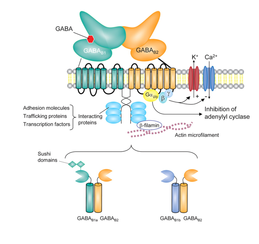
There are two types of gaba receptors, categorized as GABA-A and GABA-B receptors. GABA-A receptors are ionotropic, meaning they work via an ion channel to reduce the charge of the neuron. GABA-B receptors use G-Proteins, which instigate a series of mechanisms inside of the neuron that inevitably depresses the activity of the neuron.
GABA-A
GABA-A receptors contain what are called a ligand-gated ion channel. A lignad is simply a molecule that is capable tof binding to this receptor and stimulating it's action. Knowing this, the name is quite self explanatory. Gaba is the neurotransmitter, or ligand, that binds to the ion channel, allowing it to function. These channels are gated to negatively charged particles. When these channels open, negatively charged ions enter the neuron and the voltage of the neuron is reduced. Thus, making it much more difficult to reach the voltage required to stimulate voltage gated calcium channels and the release of extraordinary transmitters contained in vesicles.
GABA-B
GABA-B receptors are called metabotropic receptors. Metabotropic receptors do not directly open ion channels, rather than indirectly depress the neuron. These receptors work through G proteins. In short, attached to the receptor on the inside of the neuron is a G stimulatory protein. These proteins contain three different subunits, we can refer to as a,b, and y. Attached to the G stimulatory protein is a molecule of GDP which inhibits the protein. Upon binding, the GDP is released, and a different molocule called GTO takes its place. GTP is functionally the same as GDP, except it has an additional phosphate. GTP causes subunits of the G-protein to detach. The a subunit will separate from the b and y subunits, leaving an a subunit and a b/y subunit . The a subunit will then bind to another enzyme on the membrane of the neuron called adenylyl cyclase. Adenylyl cyclase is an enzyme that turns ATP from inside of the neuron into what is referred to as cAMP. The A subunit inhibits the function of this enzyme, thus levels of cAMP are reduced. For this discussion, all you need to know about cAMP is that cAMP has the ability to stimulate the activity of specific proteins involved in increasing the voltage within the neuron. Thus, inhibiting the enzyme responsible for converting ATP into cyclic amp is extremely important for inhibiting neuronal excitation.
The b/y subunit also can inhibit the neuron from reaching it's voltage threshold. The b/y subunit binds to potassium and calcium channels on the membrane of the neuron, and inhibits these channels from allowing positively charged potassium and calcium into the neuron. Plus, inhibiting this neuron from reaching its action potential.
Concluding remarks
Inhibition is a vital and necessary process to proper colander functioning. Lack of inhibition makes focusing on a singular task extremely difficult, and increases sensations of anxiety due to hyperexcitation of the brain and nervous system. It is thought that it is the tandem of an excess of glutamate and a lack of GABA that could exacerbate some of the symptoms of certain mental abnormalities , as we discussed in the above content. For individuals who may struggle with anxiety, inability to focus, and poor sleep, altering nutritional variables such as increasing dietary composition of protein and B vitamins could be a efficacious first option for helping manage some of the symptoms through the diet. That being said, consult your doctor before making any dietary, nutritional, supplemental, or medication alterations. However, I hope this review provided you with a stronger understanding of some of the amazing ways that your brain can control its activity using different neurotransmitters.
https://www.ncbi.nlm.nih.gov/pmc/articles/PMC5979278/#:~:text=Glutamine%20transporters%20on%20the%20plasma%20membranes%20of%20astrocytes%20and%20neurons,another%20round%20of%20presynaptic%20release. https://www.ncbi.nlm.nih.gov/books/NBK526124/#:~:text=GABA%20is%20formed%20from%20glutamate,post%2Dsynaptic%20terminals%20of%20neurons. https://www.ncbi.nlm.nih.gov/pmc/articles/PMC9787829/ https://www.ncbi.nlm.nih.gov/pmc/articles/PMC5153567/


